Retinal Diseases
Retinal Diseases
Diabetic Retinopathy
Diabetes and the Eye
An increasing incidence of diabetes mellitus poses a major health problem in India. The contributing factors are:
- an inappropriate diet, high in fat and carbohydrates
- sedentary lifestyle
Diabetes may affect both the young (type I) and the old (type II). The latter type is far more common. Regardless of the type of diabetes, many diabetics develop a complication called diabetic retinopathy, a change in the retinal blood vessels that leads to loss of vision.
How does diabetes affect the eye?
 Diabetes causes weakening of the blood vessels in the body. The tiny, delicate retinal blood vessels are particularly susceptible. This deterioration of retinal blood vessels, accompanied by structural changes in the retina, is termed diabetic retinopathy and will lead to loss of vision.
Diabetic retinopathy is gradual in onset and is related to the duration of diabetes. High blood glucose levels, high blood pressure and genetics influence the development and progression of diabetic retinopathy.
There are two main stages of diabetic retinopathy:
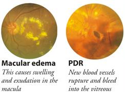
- Non-proliferative: When the blood vessels leak, macular edema may occur, thereby reducing vision.
- Proliferative: When new, weak blood vessels grow or proliferate, bleeding into the vitreous may occur and cause severe visual loss.
Eye examination in diabetic retinopathy Every diabetic is a potential candidate for diabetic retinopathy. There are no symptoms at the initial stages. Periodic eye examination with dilated pupils is the only way to detect early disease and prevent further deterioration of vision.
Diagnosis
Diagnostic tools such as a slit lamp, ultrasound and procedures such as fluorescein angiography are used in addition to an ophthalmoscope to assess whether the patient has diabetic retinopathy or other eye problems.
Fluorescein AngiographyThis is a magnified photography of the retina using an injectable dye. It helps classify the condition, record changes in the retinal blood vessels, decide on the mode of treatment and evaluate the treatment.
Treatment
Lasers are widely used in treating diabetic retinopathy. Lasers are formed by an intense and highly energetic beam of light. They can slow down or stop the progression of diabetic retinopathy and stabilise vision.
Laser and its side effectsLaser treatment is usually performed as an outpatient procedure. The patient is given topical anaesthesia to prevent any discomfort and is then positioned before a slit lamp. The ophthalmologist directs the laser beam precisely on the target with the aid of the slit lamp and a special contact lens. Absorption by the diseased tissue either seals or destroys the tissue. Additional treatment may be required according to the patient's condition.
Some patients may experience side effects after laser treatment. These are usually temporary. Possible side effects include watering eyes, mild headache, double vision, glare or blurred vision. In the event of sudden pain or vision loss, the ophthalmologist must be contacted immediately.
What is Vitrectomy?
The retina is the light-sensing tissue at the back of the eye. The vitreous is the clear, jelly-like substance that fills the middle of the eye. In some patients, there may be bleeding into the vitreous or the vitreous may pull the retina, reducing vision severely. In such instances a surgical procedure called vitrectomy is performed. The Vitreous is removed during vitrectomy surgery and usually replace by a saltwater solution.
The operation removes any blood or debris (from infection or inflammation) that may be blocking or blurring light as it focuses on the retina. Vitrectomy surgery removes scar tissue that can displace, wrinkle, or tear the retina. Vision is poor if the retina is not in its normal position. This surgery can also remove a foreign object stuck inside the eye as the result of an injury.
Age Related Macular Degeneration (ARMD)
What is macular degeneration?Macular degeneration is a condition of the eye that is often related to aging. It is commonly referred to as age-related macular degeneration, and is often abbreviated as AMD. Age related macular degeneration is the most common cause of legal blindness in the geriatric population in the west and is probably more common in India than believed.
Dry ARMD causes thinning and atrophy of the macula with variable visual loss but is not amenable to any treatments as of now. Wet ARMD results from leakage or bleeding from choroidal neovascularisation and if untreated could lead to scarring and progressive visual loss. Conventional laser therapy has been found to be effective in the management of only a selected group of patients.
Treatment
Photodynamic therapy (PDT) with Visudyne
The light sensitive drug- visudyne is injected into the patients bloodstream which accumulates in the abnormal new vessels in the eye. This drug is activated by a non-thermal laser which closes the abnormal vessels without damaging the overlying sensory retina. Studies have shown that PDT slows the progression and improves vision in some forms of the disease.
Transpupillary Thermotherapy (TTT)TTT is a cost effective alternative in the disease forms not eligible for PDT. This is a low energy diode laser which directly closes the abnormal vessels with a small risk of damage to the overlying retina.
Sub macular SurgerySub macular surgeries and macular translocation surgeries have been found to be effective in selected cases of advanced ARMD.
Source: Aravind Eye Cares
Last Modified : 2/20/2020
The topic covers various aspects of Ayurveda - bas...
This topic covers about Breast Cancer causes, symp...
This topic provides information related to the com...
This topic discuss about Acute Lymphocytic Leukemi...
